

Mitochondria are the powerhouse of the cell. They are the part of the cell responsible for turning the food into energy for the body. Mitochondrial dysfunction is linked to a number of diseases. In this article you'll learn all you need to know about the mitochondria, how they work, and how they can affect your health.
What is Mitochondria?
The mitochondrion (plural mitochondria) is a membrane-enclosed organelle responsible for the conversion of fats and carbohydrates (glucose) into usable forms of energy for the body [1, 2].
The main function of mitochondria is to produce ATP, the main source of energy used by all cells. ATP is used for all the necessary activities of the cell, and we must eat to replenish ATP production [2].
Mitochondria use oxygen to produce ATP in a process known as aerobic or cellular respiration. This reaction uses oxygen and produces carbon dioxide, which we exhale through our lungs [2].
The number of mitochondria per cell varies with every cell type. As the energy demands of a cell increase, for example in muscle cells, the number of mitochondria will also increase. This allows for more energy production and usage in active cells [2].
Aside from the production of energy, mitochondria play a vital role in monitoring calcium levels, which helps balance energy demand and production in the cell [3].
It is also responsible for initiating destruction of old and faulty cells, a process called apoptosis. This is necessary to make room for growth and regeneration of cells after injuries and to prevent cancer growth [4, 4, 5].
Byproducts of mitochondrial activities accumulate and produce free radicals. This causes oxidative stress, a leading cause of almost all age-related diseases [6].
Improper mitochondrial function leads to the buildup of waste and prevents faulty cells from being removed from the body. This can result in tumor growth and cancer [5].
Mitochondria are the target of a number of new promising therapies for cancer, heart failure, insulin resistance, and neurodegenerative disorders [5].
Since energy is central to life, improving mitochondrial function is directly related to physical and mental health.
Mitochondrial DNA
The majority of the human genome, which is all of the DNA in an individual, is packaged into 23 pairs of chromosomes in the nucleus of the cell. In addition to nuclear DNA, humans also carry a specific set of 37 genes that are only found in the mitochondrion [7].
Mitochondrial DNA (mtDNA) is a single and circular chromosome. Each mitochondrion carries 2 to 10 copies of it [8].
Mitochondrial DNA mainly codes for proteins that are needed for mitochondrial function. In contrast, the nuclear DNA codes for proteins that are required to activate the synthesis of mitochondrial proteins [8].
The 23 pairs of chromosomes in the nucleus are inherited from both parents after undergoing recombination, a process that makes you unique [9].
Conversely, mitochondrial DNA can only be inherited maternally. This means that mitochondrial DNA is passed down from the mother only. Paternal mtDNA is broken down and discarded after the sperm enters the egg [8].
Origin of the Mitochondrion
Some of the first living organisms on earth were single-celled prokaryotic organisms like bacteria. Human beings are eukaryotic organisms, meaning that we are composed of eukaryotic cells. It is hypothesized that eukaryotic cells originated from prokaryotic cells [10, 11].
Prokaryotes differ from eukaryotes in a number of ways [10]:
- Prokaryotes do not have internal compartments (organelles).
- DNA is circular, not linear like in eukaryotes.
- Prokaryotes do not have a nucleus, and thus their DNA floats freely within the center of the cell, while eukaryotic DNA is packaged into the nucleus of each cell.
- Prokaryotic DNA is simpler than eukaryotic DNA.
- Prokaryotes replicate by splitting in half and producing 2 identical copies (binary fission).
Due to their many similarities, the mitochondrion is thought to have evolved from ancient oxygen-using prokaryotic cells like bacteria [10, 11].
- Structurally, the mitochondrion resembles bacteria.
- It is composed of a double membrane through which ATP is synthesized, similar to bacterial ATP production.
- It carries its own circular DNA.
- The sequence of mitochondrial DNA strongly resembles the sequence of DNA in modern prokaryotes (Rickettsia prowazekii).
- Like prokaryotic organisms, the mitochondrion can replicate itself within the cell by binary fission.
The most widely accepted theory that explains these features is the endosymbiotic theory. This theory suggests that ancient prokaryotic organisms that produced energy-using oxygen accidentally fused with eukaryotic cells that did not use oxygen. The two cells mutually benefit from working together as an individual unit (endosymbionts) [10, 11].
The ability to use oxygen for energy proved to be favorable for both of the survival of the cells. Over time, the fused cells evolved into modern eukaryotic organisms. The mitochondrion is thought to be a remnant of the original prokaryotic organism [11].
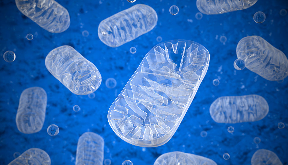
Structure
The structure of the mitochondrion is highly unusual, which is conducive to its function as an ATP-making machine [1].
The mitochondrion is enclosed by a double membrane and the inside of the organelle is called the matrix. The inner membrane folds multiple times into structures called cristae. The ATP-making proteins are located between these two membranes and movement of ions across the inner membrane powers these proteins. The presence of the cristae increases the surface area available for ATP production [1, 3].
The matrix contains a copy of the circular DNA and proteins responsible for mediating mitochondrial activity. These include proteins that create ATP, stimulate mitochondrial division and the fusion of mitochondria, metabolize molecules, and induce apoptosis [1].
Mitochondria can undergo division (fission) and fusion. Mitochondrial division increases the number of mitochondria by dividing the mitochondrion in half or producing a small fragment of the original mitochondrion [3].
Mitochondrial fusion occurs when two mitochondrion fuse together, forming an elongated structure. This usually occurs when there is damage. By fusing together, mitochondria can overcome the dysfunction allowing the two semi-damaged mitochondria to work as one fully-functioning mitochondrion (complementation) [3].
Mitochondria Vs. Chloroplasts
The structure and function of mitochondria are very similar to chloroplasts. Chloroplasts are found within plants and some algae. Like mitochondria, they:
- have their own double membrane
- have their own set of DNA and RNA
- provide energy to the cell
- have similar sets of enzymes and proteins
Unlike mitochondria, chloroplasts use sunlight to make energy for the plant through a process known as photosynthesis.
Mitochondria Function
The mitochondrion is responsible for the conversion of fats and carbohydrates into ATP, the energy currency of the cell. In addition, mitochondria can induce cell death when necessary, manage calcium levels, and break down carbon-based molecules [3, 12].
Byproducts of normal mitochondrial function accumulate and produce free radicals. This leads to oxidative stress, which is a leading cause of almost every serious age-related disease [12].
1) Mitochondria Are the Powerhouse of the Cell
The most significant role that mitochondria play is the production of ATP. A series of biochemical reactions both inside and outside of the mitochondria results in the production of ATP through a process called oxidative phosphorylation. Aside from having a normal cellular function, these reactions require food and oxygen for energy production [13].
Fats, carbohydrates, and proteins all provide the body with different amounts of ATP when broken down [12].
Carbohydrates, which are converted to glucose molecules, are the quickest to break down and generate 34 to 38 molecules of ATP for every molecule of glucose that undergoes respiration. They are the body’s preferred form of energy [14].
Fats are used for energy when glucose levels are low. A single molecule of palmitic acid, a common saturated fat, can produce 130 molecules of ATP. However, the breakdown of fats (lipolysis) is a much more complex process, and it can take up to 72 hours to digest a fatty meal [15].
Proteins are only used for energy when the body is starved [15].
What Is Cellular Respiration?
There are 3 stages in cellular respiration [16]: glycolysis, Krebs cycle, and oxidative phosphorylation.
Glycolysis
Once carbohydrates or glycerol from fats is broken down into glucose molecules, they undergo a series of reactions called glycolysis [3].
- Glycolysis occurs outside of the mitochondria, in the cytosol of the cell.
- It is an anaerobic process, meaning that it does not need oxygen to occur.
- It results in the net production of 2 molecules of ATP, 2 molecules of NADH+, and 2 molecules of pyruvate.
Glycolysis alone does not generate enough energy for the cell, but the products of glycolysis travel into the mitochondria where they pass through the Krebs cycle, also known as the tricarboxylic-acid cycle (TCA) or the citric acid cycle [3].
- Pyruvate and NADH diffuse into the mitochondria (matrix).
- Pyruvate undergoes a reaction that generates 2 intermediary molecules called acetyl-CoA.
- Carbon dioxide is produced as a waste product and can be excreted through the lungs.
Fats are converted into glycerol and fatty acid tails. Glycerol can be broken down into glucose in the cytosol, which then enters the cellular respiration pathway at glycolysis. The fatty acid tails undergo a series of reactions through a process called beta-oxidation. This produces acetyl-CoA, which can enter respiration at the second stage [3].
In the event of starvation (absence of both carbohydrates and fats), amino acids from proteins are converted to small carbon-based molecules in the mitochondria. These molecules can enter the respiratory pathway at the Krebs cycle to produce ATP. This is harmful because the proteins used to provide energy are taken from our bones, muscle, and skin [16].
Krebs Cycle
The Krebs cycle is the stage in which the glucose is completely converted to carbon dioxide using oxygen (oxidation). It involves a series of reactions using the acetyl-CoA previously generated. Acetyl-CoA produced from the breakdown of fats enters respiration at this stage. The objective of the Krebs cycle is to produce high-energy molecules that can later be used to generate a larger amount of ATP [14].
- Acetyl-CoA enters the Krebs cycle in the mitochondrial matrix.
- Four reactions requiring oxygen result in the net yield of 2 ATP, 6 NADH, and 2 FADH2 molecules.
NADH and FADH2 are high-energy molecules that provide the greatest source of energy for ATP production. They are used in the third stage of respiration, oxidative phosphorylation [14].
Oxidative Phosphorylation
This stage of respiration is the most important as it generates the largest amount of ATP. This is where the structure of the mitochondrion plays a large role. Also, this process is the reason we need oxygen to survive [17, 3].
Between the outer and inner membranes of the mitochondrion exists a small space called the intermembrane space. A group of mitochondrial enzymes and protein complexes line the inner membrane, forming a structure called the electron transport chain. Oxidative phosphorylation occurs in two steps: electron transport and chemiosmosis [17].
Electron Transport Chain [17]:
- Six NADH and two FADH2 undergo chemical reactions and donate their electrons to the first of the protein complexes in the electron transport chain.
- NADH becomes NAD+.
- FADH2 becomes FAD.
- Electrons are passed from complex to complex along the chain.
- As electrons pass from one protein complex to the next, they generate energy.
Chemiosmosis [17]:
- The energy generated by passing electrons down the transport chain is used to pump H+ ions from the inner mitochondrial matrix into the intermembrane space.
- The H+ ions accumulate between the two mitochondrial membranes.
- At some point, the amount of H+ ions within the inner membrane space is much larger than the amount of H+ within the mitochondrial matrix, creating a chemical gradient across the inner membrane.
- H+ ions flow back into the matrix down their concentration gradient, releasing a large amount of energy.
- The final enzyme of the electron transport chain, ATP synthase, harvests the energy generated by the flow of H+ ions into the matrix.
- ATP synthase uses this energy to generate 34 to 38 ATP molecules and 6 H2O molecules.
The purpose of oxygen in the respiratory process is to pick up the H+ ions that flow back into the cell and form water molecules. If we had no oxygen, the H+ ions would remain within the mitochondrial matrix. This would diminish the gradient created across the inner membrane because there will be H+ ions on both sides of the membrane. This will prevent chemiosmosis and ultimately, there will be no energy for ATP production [17, 3].
2) Mitochondria Can Trigger Cell Death
Mitochondria play a major role in initiating cell death when the cell is either damaged or aged. This is useful to focus the energy production on cells and tissues that have higher energy demands. Cellular death also prevents the spread of mutated and faulty cells. The process of cellular suicide is called apoptosis, which is largely mediated by the release of “suicide” proteins from the mitochondria [4, 18].
When only a mitochondrion is damaged, mitochondria can destroy themselves, by a process called mitophagy, leaving the cell and other mitochondria intact [18].
The electron transport chain, found in the inner membrane of the mitochondrion, often leaks free radicals, or reactive oxygen species (ROS). While free radical production is normal, it can lead to harmful oxidative stress in the cell. Free radicals react with many other substances in the cell, leading to DNA damage and destruction of many vital proteins [4, 18].
The buildup of oxidative stress is linked to over 200 human diseases. Apoptosis, or programmed cell death, prevent the accumulation of free radicals. Mitochondria can respond to apoptotic signals inside (intrinsic pathway) and outside of the cell (extrinsic pathway) [4, 18].
Aside from free radicals, certain proteins can bind to receptors on the surface of the cell and initiate apoptosis. These are called death signals, and they are released in response to DNA mutations, radiation, or starvation of nutrients [4, 18].
When death signals bind to their receptors on the cell, they cause a number of chemical reactions that lead to changes in the mitochondrial membrane. This leads to the movement of proteins into the mitochondrial matrix, releasing a protein called cytochrome C from the inner membrane [4, 18].
Cytochrome C is one of the proteins in the electron transport chain. When released from the mitochondrion, the cell must undergo apoptosis. It is the determining factor of cell death [3, 18].
The destructive events of apoptosis are carried out by caspase enzymes. Caspases exist in their inactive form in every cell. When cytochrome C is released, a series of chemical reactions activate the caspase enzymes. Caspases cleave, or cut up, the different proteins in the cell. As the process goes on, the caspases lead to complete destruction of the cellular contents [18].
3) Mitochondria Plays a Central Role in Breaking Down Most Organic Molecules
The mitochondria contain enzymes that break down many different molecules to be used for energy in the cellular respiration pathway. These enzymes also produce intermediary molecules that are made up of only one carbon atom. These 1-carbon-based molecules are highly reactive and can interfere with natural cellular activities [19, 20].
Inside mitochondria, 1-carbon molecules are converted into different amino acids that are needed for ATP and protein production. Vitamins B9, B12, B6, and B2 provide the carbons needed to carry out these reactions. Deficiencies in B vitamins are linked to age-related diseases like Alzheimer’s disease, cardiovascular disease, and cancer [19, 21].
The production of haem groups, the structural core of hemoglobin, found in red blood cells, is an example of one important substance produced by 1-carbon metabolism in the mitochondria. Hemoglobin is an iron-containing protein that allows for oxygen and carbon dioxide to be carried in the blood and transported around the body [12].
The 1-carbon-based molecules are exported from the mitochondria to produce some of the building blocks of DNA. This occurs in the jelly-like fluid that fills the inside of a cell (cytoplasm), where the products can either be moved to different parts of the cell as needed or exported to other tissues [21].
The mitochondria also form part of the urea cycle, the process in which nitrogen atoms from different molecules are excreted through the body as urea. Urea is the main substance found in urine, producing its yellow color [12, 22].
Therefore, the mitochondria prevent the disruption of normal cellular function, participate in the removal of waste products, and provide precursor molecules needed for other reactions.
4) Mitochondria Balance Calcium Levels
Mitochondria monitor calcium levels in the cell to coordinate energy production with energy demand. Calcium levels increase in the cell for the activation of almost any biochemical pathway. Activating a cell, in turn, requires energy, so mitochondria will have to increase ATP production [3].
When calcium enters the cell, the concentration of calcium increases outside of the mitochondrial matrix. This creates a concentration gradient that drives the flow of calcium into the matrix. Protein channels that allow calcium entry is located on the inner membrane and open in response to high intracellular calcium concentrations [3].
In muscle cells, calcium entry results in increased mitochondrial production. This allows for the energy supply to meet the energy demand of that tissue [3].
Diseases Linked to Mitochondrial Dysfunction
Properly functioning mitochondria are central to health, as it is the main energy provider of the cell. However, reactive oxidative species produced by mitochondria accumulate over time, and oxidative stress leads to age-related diseases. Due to the vast role of mitochondria in the cell, mitochondrial dysfunction is linked to hundreds of diseases [5, 23, 24].
Additionally, a number of metabolic disorders are caused by genetic mutations in either mitochondrial or nuclear DNA. These mutations may be inherited or occur randomly [23].
1) Cancer and Mitochondrial Dysfunction
Cancer cells require mitochondria to power the growth of tumors. Cancer cells have an increased number of mitochondria to provide this energy. The mitochondrial increase is mediated independently by different transcription factors or proteins that initiate the production of specific genes [5].
On the other hand, cancer cells increase the turnover of mitochondria that have accumulated free radicals. Oxidative stress is increased in cancer cells, which damages the surrounding tissue [25].
One of the hallmarks of cancer is the ability of the cell to avoid programmed cell death (apoptosis). Normally, the mitochondria of healthy cells would trigger this process if the cell was replicating too much or too quickly. However, in cancer cells, programmed cell death is avoided by increasing the destruction of mitochondria that have accumulated free radicals. They also turn on antioxidant pathways so that oxidative stress does not trigger cell death [25, 5].
Mitochondria of cancer cells have lower levels of proteins that promote cell death (BAX/BAK) and/or higher levels of proteins that prevent cell death (BCL-2/BCL-XL) [25, 5].
The network of mitochondria in cancer cells is also different. Cancer cells have more fragmented mitochondria (by increasing mitochondrial division and decreasing fusion) [25].
Cancer cells also produce energy differently through a process known as the Warburg effect. Energy production is largely done without oxygen (anaerobically), via glycolysis. This may be caused by turning down mitochondrial function to avoid apoptosis [25, 5].
Aerobic respiration through the Krebs cycle and oxidative phosphorylation still occur in cancer cells but to a lesser degree.
2) Neurodegenerative Diseases and Mitochondrial Dysfunction
Mitochondrial dysfunction is a large cause of age-related neurodegenerative diseases like Alzheimer’s, ALS, and Parkinson’s disease. The accumulation of free radicals with age results in the DNA and protein damage. Mitochondria accumulate defective proteins that cause loss of energy production and ultimately cell death [24].
Alzheimer’s Disease
- Amyloid beta proteins build up around the outer mitochondrial membrane.
- This buildup decreases ATP production, increases oxidative stress, and ultimately causes cell death.
- Amyloid beta increases mitochondrial protein production.
- Mitochondrial enzymes have decreased activity, leading to reduced ATP levels.
- Mitochondria undergo structural changes within the cell. Rather than existing in long tubes (mitochondrial fusion), the mitochondria are fragmented into little pieces within the cell (fission). This adds to the overall dysfunction of brain cells seen in patients with Alzheimer’s disease [24].
Parkinson’s Disease
- The hallmark of Parkinson’s is the accumulation of alpha-synuclein protein, leading to cell death and loss of neurons.
- Patients with Parkinson’s accumulate this protein in the mitochondria, leading to increased oxidative stress and reduced energy production.
- Parkin protein (E3 ubiquitin ligase) and PINK1 protein are responsible for marking damaged mitochondria for destruction. Patients with Parkinson’s disease have low levels of these proteins.
- Damaged, low functioning mitochondria are not degraded and remain in the cell.
- Alpha-synuclein continues to accumulate, leading to neurodegeneration [26].
3) Diabetes and Mitochondrial Dysfunction
Improper mitochondrial function is seen in patients with both type 1 and types 2 diabetes.
Aside from having a lack of glucose for respiration, the network and shape of mitochondria in the cells of diabetic patients are abnormal [27, 5].
Mitochondria are broken up into small fragmented networks (increased division, decreased fusion) throughout the cell. This is seen in both type 1 and type 2 diabetes [27].
Type 2 diabetes is caused by insulin resistance in cells, reducing the amount of glucose available for respiration. This decreases ATP production and thus the energy available to the cell [27].
It is unknown whether mitochondrial dysfunction is a cause of insulin resistance or a symptom of it. Some evidence points to the mitochondria as a cause of insulin resistance, rather than a symptom [27].
Patients with type 2 diabetes have reduced levels of mitochondrial proteins responsible for energy production [27, 5].
4) Heart Failure and Mitochondrial Dysfunction
Heart cells rely heavily on mitochondria to power the pumping of the heart. Mitochondrial dysfunction is implicated in heart failure due to the buildup of oxidative stress [5].
Patients with heart failure exhibit reduced mitochondrial activity. The mitochondria have lower activity at electron transport chains. This is caused by a loss of oxygen supply to the mitochondria [5].
Since oxygen supply is reduced, electrons at the electron transport chain cannot be picked up by oxygen. This leads to the accumulation of electrons, which produce free radicals [5].
5) Chronic Fatigue Syndrome and Mitochondrial Dysfunction
Chronic Fatigue Syndrome, commonly known as CFS, is a lifelong disorder characterized by prolonged (>6 months) symptoms of intense fatigue that can reduce daily functioning by over 50%. Patients with CFS suffer from a variety of other symptoms including [28]:
- Anxiety
- Depression
- Headaches
- Muscle Pain
- Cognitive Dysfunction
Although it was once thought to be a disease in the mind, increasing evidence points to mitochondrial dysfunction as one of the leading causes of this disorder [29].
Multiple clinical trials have been conducted and have produced mixed results. CFS may be caused by one or more of the following mitochondrial abnormalities [R]:
- Smaller mitochondrial shape and number
- Lower L-carnitine, ALCAR, ubiquinone or CoQ10 levels
- Reduced protein activity in the electron transport chain (oxidative phosphorylation)
- Reduced ATP production
A number of studies indicated that there were no significant differences in mitochondrial structure or function of healthy and normal patients. Further studies are required to fully understand the cause of this condition, the role of mitochondrial dysfunction and how to effectively treat patients.
6) Genetic Mitochondrial Disorders
Genetic mutations in mitochondrial genes can result in mitochondrial dysfunction through 1 or more of 5 distinct mechanisms [23]:
- Inability to utilize other molecules (substrates) for energy production
- Improper Krebs cycle
- Defective energy production through the electron transport chain (oxidative phosphorylation)
- Defective transport of molecules in and out of the mitochondria
- Defective proteins in the electron transport chain
These defects can be caused by mutations in mitochondrial and/or nuclear DNA. Additionally, defects can occur when mitochondrial DNA is unable to communicate with nuclear DNA [23].
Some metabolic diseases that are caused by mitochondrial and/or nuclear DNA mutations include [23, 5]:
- mtDNA depletion syndrome (MDS): MDS refers to a group of disorders, any of which have dysfunctional mitochondrial DNA. This can result in different developmental, muscle, and brain abnormalities. Many known mitochondrial diseases are a result of MDS.
- Mitochondrial myopathy: a Mitochondrial disease that causes muscle problems such as weakness, exercise intolerance, breathing difficulties, or issues with vision.
- Mitochondrial encephalomyopathy, lactic acidosis, and stroke-like episodes (MELAS): A mitochondrial disorder that affects the brain and muscle throughout the body. Stroke-like episodes and the buildup of lactic acid results in dementia, vomiting, extreme pain, and muscle weakness.
- CoQ10 deficiency: A deficiency in coenzyme Q10, a protein that is part of the electron transport chain.
- Mitochondrial neurogastrointestinal encephalomyopathy (MNGIE): A rare mitochondrial disease that primarily affects the brain and digestive system. The muscles and nerves of the digestive system do not properly push food through the system.
- Mitochondrial diabetes: Referred to as maternally inherited diabetes and deafness (MIDD), a subtype of diabetes caused by a single mutation in the mitochondrial DNA (at position 3243). The disease results in loss of hearing and diabetes similar to type 1.
- POLG mutations: A gene that codes for DNA polymerase subunit gamma, the active (catalytic) part of the mitochondrial protein responsible for DNA synthesis. Mutations in this gene lead to the defective production of mitochondrial proteins and many mitochondrial diseases.
The Mitochondrial Bottleneck Effect
Carrying mutations in your mitochondrial DNA does not necessarily mean that you will transmit the disease. The proportion of cells carrying mutated mitochondria has to be more than the cells carrying healthy mitochondria in order for the disease to be seen [30].
During female egg cell (oocyte) production, each egg cell will carry a random selection of mitochondrial DNA copies. Some of the copies may carry mutations, whereas some may be completely normal. As the egg cell matures and prepares for fertilization, many mitochondria are replicated at random. This distributes the chance of inheriting mutant mitochondria [30].
This phenomenon means that if the mother carries highly mutant mitochondria, her offspring will not necessarily carry the trait [30].
If enough cells carry mutant mitochondrial DNA, for example, more than 50% of the cells in the body, it is likely that the child will have the associated disorder.
Preventing Inheritance of Dysfunctional Mitochondrial DNA (mtDNA)
New technologies can prevent the transmission of mutated mitochondrial DNA from the mother to the offspring. A new technique, called the 3-parent in vitro fertilization, consists of switching nuclear DNA from the mother with the nuclear DNA of a donor female egg that has healthy mitochondrial DNA. Therefore, the donor egg carries the genetic information from the mother but lacks the mutated mitochondrial DNA that she carries as well.
Using in vitro fertilization, the egg is then artificially inseminated using the paternal semen. The fertilized egg is then reintroduced into the mother’s uterus, where it can latch on to the uterus [31].
The offspring will have all of the physical characteristics of their biological parents because the nuclear DNA is unchanged. The only thing that is different is that this child will have a proper mitochondrial function, unlike the mother who carries mutated copies of the mitochondrial DNA [31].
You May Have Mitochondrial Dysfunction if:
- You feel excessively tired [32]
- You are unable to exercise for long periods of time [33]
- You feel short of breath, especially during exercise [34, 35]
- You have poor bone growth and health [36]
- You have difficulty controlling your movements, balance, and coordination (ataxia) [37]
- You have trouble walking or talking [38]
- You have muscle weakness and pain [39]
- You have heart muscle disease (cardiomyopathy) [40]
- You have a gut and digestive issues [41]
- You have liver and kidney disease [42]
- You have droopy eyelids, vision loss, and other eye problems [43]
- You have diabetes and other hormonal disorders [27, 44]
- You have trouble hearing [45]
- You experience more migraines, strokes, and seizures [46, 47]
- You have difficulty remembering things [47]
- You have a developmental delay [48]
- You are autistic [49]
- You have recurrent infections [50, 51]
